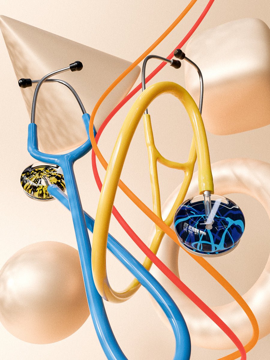
A stethoscope is a basic medical instrument used by practitioners in the medical field to listen to internal organs' sounds. It is generally used for diagnosis, as the physician can listen to sounds picked and interpret various medical conditions for treatment.
A stethoscope primarily consists of a disc-shaped resonator, a chest piece placed on the skin to pick sounds, two tubes that connect the earpiece for sound transfer, and the earpiece to listen to sounds captured for diagnosis. These parts work together for accurate sound capturing and efficient transfer when connected properly. You may also want to read how to properly wear your stethoscope as well.
Even though the stethoscope is a common instrument, there are several interesting facts you need to know about it. Follow this guide to know more about a stethoscope as we highlight the top ten facts for this diagnostic instrument.
1. A French man invented it
Doctor Rene Theophile Hyacinthe Laennec, a medical cadet in the French revolution, was the first man to invent and use a stethoscope in 1816. Before this invention, doctors diagnosed patients by placing their hands against the chest to feel vibration or heartbeat, thus picking basic sounds, though they would miss quieter ones.
The prominent diagnostic technique was to press the ear on the patient's chest to pick sounds and again, this was inefficient as there was no amplification. This had another major limitation: male doctors could not feel comfortable pressing their ears against their female patients' chest area.
This major limitation is what led to the invention of the stethoscope.
In 1801, Doctor Laennec moved to Paris to finish his medical studies and in 1816, he made this invention when examining his female patient. It was awkward for him to place his ear on the patient's ample breast and he also found it improper.
Remembering the acoustic effect of hearing a pin scratch a piece of wood while listening on the other side and the natural sound amplification involved, made him act swiftly. He rolled a piece of paper, placed it on the patient's chest and he could hear the heart and lung sounds more clearly. Thus he concluded that a tube would work better compared to an ear alone.
After this incident, Doctor Laennec went ahead to design a hollow wooden tube with an earpiece on one side and a flared end for this purpose. He then named this instrument a stethoscope, a two Greek word stethos and skope meaning chest and examination, respectively.
2. It was the first effective non-invasive tool
Before the invention of a stethoscope, there was no efficient non-invasive diagnostic technique as the tools were unreliable and poor. The doctors had no faith in the non-invasive techniques as they often resulted in misdiagnosis. Stethoscope's high accuracy in the diagnosis of respiratory conditions made it the first effective non-invasive tool.
3. There are two types of stethoscope, monaural and binaural
Initially, doctors used the monaural stethoscope (one earpiece), but with evolution, a binaural stethoscope (two earpieces) was made and the latter is commonly used today due to its high efficiency.
The stethoscope developed by Doctor Laennec was monaural as it had one earpiece and this instrument caught on as the majority of the doctors used it. After ten years, in 1828, Adolphe Piorry designed a more flexible stethoscope using simpler and less awkward devices though still, it was monaural.
In 1852, the first binaural stethoscope was developed by George Cammonn, a New York physician. His version included two ivory earpieces connected to two tubes covered in silk by a hinge with a bell-shaped listening device. This binaural then evolved into modern stethoscopes types, now available with very high efficiency.
Upgraded monaural stethoscopes are used today to listen to unborn babies' heartbeats through a mother's skin.
4. It has three parts
A stethoscope generally contains three main parts the headpiece, tubing, and chest piece.
Headpiece
The headpiece is the half upper part of the stethoscope containing ear tubes, a metallic section connecting ear tips, and the tubing. Ear tubes separate sound into right and left channels for perfect hearing and they are designed to isolate and transmit sound with minimal loss.
The second part is the tension springs which adjust the headset tension by pulling the ear tubes to reduce tension and squeezing them together to increase tension. The last part of the headset is the ear tips made of silicone or rubber, which go to the user's ears where they receive sound from the earpiece.
Tubing
This is the soft, flexible line used to make sure sound transferred is of the same quality and frequency as that captured by the diaphragm and bell. It connects the stem of the heart-piece.
Chest-piece
The chest piece contains the stem, diaphragm, and bell. The stem connects the tubing and the chest piece. Other than connecting the two, it allows the user to switch between the chest-pieces, diaphragm, and bell. The diaphragm and bell are used to pick sounds by placing them on the patient's skin and then transferring the sound to the earpiece. However, advanced ones can still pick sound over thin clothing.
5. The chest part can have a bell or diaphragm
The chest part, which is the chest-piece, can have a dual head having both the diaphragm and a bell or, in other cases, just one for the single head. The diaphragm is the large circular end used to capture higher frequency sounds as it covers a wider area. On the other hand, the bell is a small circular end that captures low-frequency sounds impossible to capture with the diaphragm.
Both are covered with non-chill and hypoallergenic material for maximum patient comfort.
6. The headset follows human ear anatomy
The earpieces are designed to be worn angled forward towards the nose to align them with the ear canal. The binaural is designed to point away from the user since the ear canals face the face and not backward or sideways. In case you wear the earpiece facing backward, you will not hear anything when using the stethoscope.
7. Uses sound to diagnose
A stethoscope basically uses sound for diagnosis; the doctor places the chest-piece against the patient skin and then listens to sounds from the heart, lung, and stomach areas. The diaphragm and bell pick high frequency and low-frequency sounds, respectively, and then transfers them to the earpiece through the tubing. The doctor carefully listens to the transmitted sounds and then evaluates them for accurate diagnosis followed by apt treatment.
8. They are used extensively for heart examinations
A "lub dub" noise characterizes heart valves' opening and closing. Doctors use the stethoscope to evaluate the valve and heart health by listening to these sounds. They also listen to heartbeats for heart health evaluation and since there are few techniques for heart evaluation, a stethoscope is commonly used.
9. Easily help diagnose respiratory illnesses
Any upper respiratory illness is easily diagnosable using a stethoscope as the stethoscope can listen to patients' breathing. The doctor can identify normal and abnormal sounds through respiratory auscultation, thus diagnosing the respective respiratory condition.
10. They are sanitized using UV light
The stethoscopes work by placing them against the patient's skin and here, they easily pick bacteria and germs. To prevent the transfer of bacteria and germs from one patient to another, the doctors sanitize them using UV radiation. UV radiation is very effective as it kills all germs and bacteria.
You can also use alcohol to clean the stethoscope. For the ear tips, pull them off them, then clean with alcohol-dipped cotton. For diaphragm and bell, do the same but clean all crevices and after cleaning, allow the parts to dry before connecting them back.
Conclusion
Hopefully, you have known more about a stethoscope, a valuable medical device, from the facts discussed above. At least you now understand how it was invented/ evolved and some interesting aspects about its general operation. We are certain that next time you will use this crucial instrument, this information will sink and help you appreciate the work that was done to produce the highly effective piece of medical equipment that today you and your patients greatly rely on.


